[mashshare]
Jurdan Mendiguchia is the Director of ZENTRUM Rehab & Performance Center, as well as a sports physical therapist working in a high elite environment (soccer, basketball, track and field, football, etc.). He consults on rehab and injury prevention for soccer (Europe), and NBA, NFL, and track and field athletes and teams. Additionally, Mendiguchia is a lecturer with some clinical research manuscripts published mainly around hamstring injury.
Freelap USA: The hamstring has received a lot of attention recently with discussions on sonography. Could you explain why other muscle groups may need attention? For example, the adductor magnus and hip flexors are also part of the equation, but we don’t see testing equipment or sonography on those muscles.
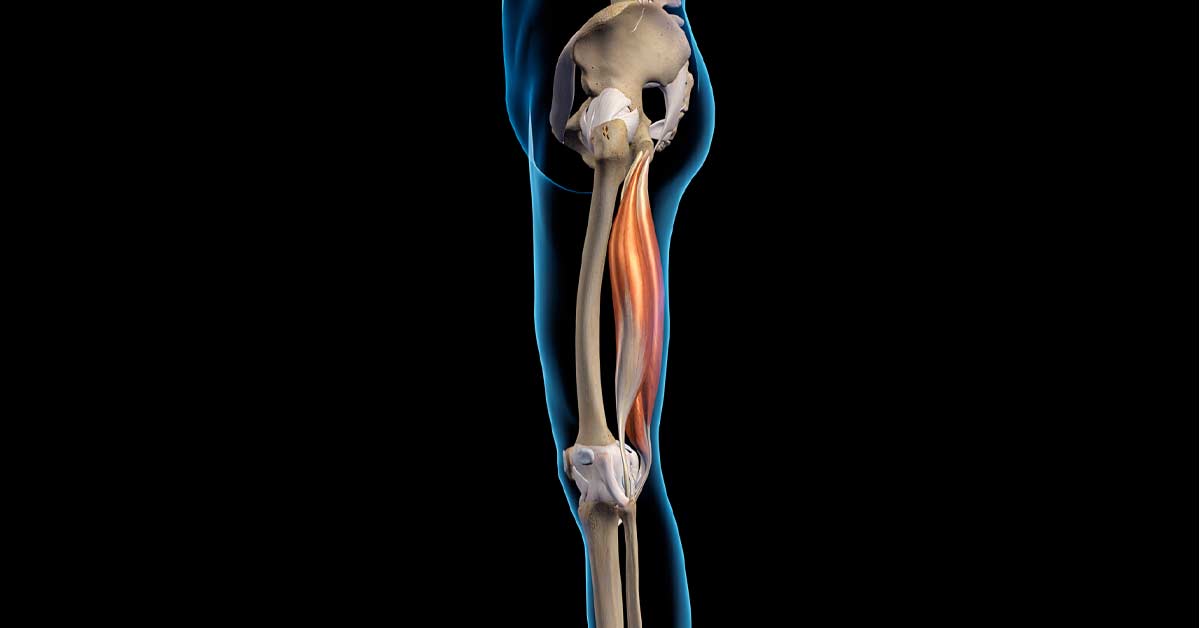
Jurdan Mendiguchia: Historically, we are used to focusing on and treating the exact injury location, but for example, what happens at the same time or concomitantly to the hypothetical moment where the injury occurs during the late swing phase of sprinting? The timing of the maximum biceps femoris length was synchronous with peak biceps femoris and gluteus maximus forces, contralateral iliopsoas peak length and forces, and also that of the peak pelvic anterior tilt. Therefore, if we assume strain as the major determinant of tissue failure, the peak length of the biceps femoris during the late swing phase of sprinting appeared to be influenced by the actions of the muscles crossing the hip joint as well as by the pelvic anterior tilt.
In addition, we know that a lot of elastic energy is transported from one leg to the other each step, and this occurs 4–5 times per second during sprinting. As far as we know, the pelvis is the unique joint that links both legs, and therefore the pelvis is the key anatomical lever and energy transfer structure between the two limbs.
Similarly, in the overstretching or kicking type of injury, a change of trunk flexion and/or anterior pelvic position would translate the ischial tuberosity superiorly (origin of the hamstring musculature), resulting in a greater active lengthening and passive tension demand of the hamstring due to a greater moment arm derived from the relative hip flexion.
Different models have also shown the influence of different muscles (adductor magnus, erector spinae, internal oblique, iliopsoas) on the length of the femoral biceps. Some of them, as you mentioned, like the adductor magnus and contralateral iliopsoas, reach magnitudes similar to those that would result in altering the biceps itself during sprinting.
In summary, you can manipulate and screen the result of the equation (biceps femoris) and, in addition, you can alter the causative factors that impact the results (both biceps femoris length and force). Our vision is to act on the whole picture, but specifically on the factor that we believe decisively alters and influences our athlete in a specific and individual way.
With regard to the study of the architecture of the biceps femoris from ultrasound measurements, it is again necessary to look at the RESULT of the equation, but I also believe that a very simplistic and superficial study of the architecture has been made comparing it with the great advances described lately in the muscle physiology area. It is surely due to the technical limitations derived from the static and reduced capability of field of view of the current existing technology, where fascicles, aponeurosis, curvature, etc. were ESTIMATED. Assuming that it is a static and local measurement (a hamstring muscle architecture change along the muscle), HOW can we predict what will happen dynamically in the architecture during sprinting without considering the effect of other muscles (iliopsoas, abdominal muscles), the pelvis, muscle tendon interaction and behavior, muscle shape change during contraction, etc.?
Also, the addition of sarcomere in series has been suggested as the phenomenon that explains the increase in fascicle length after eccentric exercise and protects from muscle damage. However, in the first unique human experiment measuring fascicle length tension rather than joint angle torque, the authors observed that protection from a repeated bout of eccentric exercise was conferred without changes in muscle fascicle strain behavior, and they suggested connective tissue structures, such as extracellular matrix remodeling, are a cause of the protective effect.
Moreover, I will tell you one last thing. The data I have, together with my colleagues Antonio Morales Artacho and Gael Guillhem—both great muscle physiologists working at INSEP in Paris—which is made with the most advanced imaging techniques today, does NOT show changes in static or dynamic fascicle level or tendon after several weeks of eccentric training protocol. Therefore, I believe that we must at least be cautious, given the technology limitations we have, when associating fascicle length as the reason for the success of eccentric exercise to prevent hamstring injuries.
Given our technology limitations, we must at least be cautious when associating fascicle length as the reason for the success of eccentric exercise to prevent hamstring injuries. Share on XFreelap USA: Postural changes to soccer players during sprinting is a bold claim. Can you explain how valuable pelvic control during sprinting is for those who are trying to address hamstring injuries? Those with anterior tilt may not always get hurt, but if they do have stride changes that increase the swing phase, they could be susceptible to injury.
Jurdan Mendiguchia: That’s a very good question, but I am unaware of an intervention study showing that ONLY pelvic tilt change was able to reduce hamstring injuries. We are right now trying to address this topic, but inside an individualized multifactorial prevention approach in a professional soccer prevention research project. There are some soccer and baseball studies showing a prospective association between pelvic tilt and hamstring tears. As every risk factor is a part of the puzzle, it will probably be beneficial for those who have excessive pelvic tilt. There is evidence that soccer players, compared to other sport athletes, showed an increased anterior pelvic tilt probably due to the type of sport requirements.
But, first of all, you would have to ask yourself: Are we able to alter the position of the pelvis? Until today, and even if the pelvic position is taken for granted, there is no intervention that shows that we can change it during high speed. Here is where I can help you today—we are close to submitting for the first time an article where we were able to decrease pelvic tilt (an average of 5–6 degrees during late swing phase) at top speed after six weeks of a multimodal intervention.
Bearing in mind that during the maximum velocity phase the biceps strain increases, especially at the proximal level, reducing the supero-anterior migration of the ischial tuberosity (hamstring muscle insertion) seems likely to decrease the strain of the biceps femoris, considered the major determinant of tissue failure. Therefore, the evaluation can be an interesting tool for those whose anteversion can be a risk factor. We also found other kinematic changes related to performance improvement, according to the world’s best track and field coaches actually, so we would act again on the PREPARE AND REPAIR concept.
Freelap USA: Manual therapy is hard to quantify but many athletes who are involved with sprinting or running may have overactive erectors. Can you explain the value of combining the relaxation of those muscles with strengthening the internal oblique?
Jurdan Mendiguchia: Among other things, we took into account such ideas when designing the proposed multimodal program to try to change pelvis position. A greater cross-sectional area (CSA) of the erector spinae has been demonstrated in sprinters as well as after a football season, probably associated with its extension function during the sprint propulsion phase (backward thrust). Because of its lever arm, as well as the increase in EMG associated with the anterior pelvic tilt, the erectors can be a solution to take care of other muscle groups.
Since influence on the biceps strain during sprinting has also been seen in models, we can conclude that multiple variables matter in injury patterns. There are studies where lumbar manual techniques have influenced the neural and muscle extensibility of the hamstring muscle group in elite male soccer players. On the contrary, the internal oblique has been associated with a decrease in biceps strain in modeling studies, and we have new data that shows a high association in an unanticipated task between the EMG of the internal oblique and the pelvic tilt.
Anyway, I do not know if we can be so specific to an actual single muscle and if it is even worth it. However, I can tell you that a certain intervention program is capable of influencing the position of the pelvis, and we have taken into account the different functions of the muscles as well as the adaptations that football generates in that musculature when designing our program.
Freelap USA: You have developed some world-class algorithms for return to play, yet we still have teams using a simple recipe of steps based on weeks rather than outcomes. Can you explain how a team can create decision-making trees or rehabilitation algorithms more successfully?
Jurdan Mendiguchia: Unfortunately, in my experience at least, there are no magic recipes here. I believe that the art lies in finding what causes your athlete to get injured—find the cause and try to modify it. If you always use a general tool or aspirin for everyone, I think the chances of success decrease. In fact, assuming that it is a multifactorial injury, the use of a single strategy does not seem to make much sense.
In summary, we will need a complete screening that includes the structural individuality of the athlete with the different risk factors (how they interact and which one influences more or determines the other) contextualized to the injury mechanism, as well as to the sport that they practice, in order to prescribe and design a rehabilitation program according to their needs and context. This process will allow clinicians to assign more importance to one thing or another depending on the characteristics of the athlete they are treating during the rehabilitation process.
If we continue using the simplistic single joint torque or isolated flexibility assessment that isn’t related to what is happening in the main injury mechanics, we will miss many things. Share on XIf we continue with the simplistic single joint torque or isolated flexibility assessment (e.g., AKE test) that is not related to what is happening in the main injury mechanics, I think we will miss many things. For me, that is the challenge of hamstring science. Provide resources and tools to be able to individualize the clinic on a day-to-day basis and know which of the tools to use with the athlete (individual and different) who is in front of you.
Freelap USA: Many sports medicine professionals don’t use video analysis to help athletes. Could you explain how video assists in the rehabilitation process? Perhaps you can go into contact times and leg recovery mechanics in detail?
Jurdan Mendiguchia: I think that video analysis in the near term can be a very useful tool for both rehab and prevention. As I have been saying, it’s one more piece of the puzzle, but it needs to be monitored because sprinting represents the main injury mechanism and both mechanics and technique should be addressed. Soon our group will publish how video analysis, in a simple and 2-D way, can indirectly give us information about the way our athletes run. As the sprint is a sequence of movements where one depends on the previous one, by extracting two key points we can deduce specific problems.
For example, let’s talk about the famous “backside mechanics” characterized by an overextended lower back (excessive arching) and/or emphasizing anterior pelvic tilt heading into touch-down. Excessive backside mechanics cause the trail leg to swing all over the place behind the center of mass. Too much forward leaning throughout the sprint cycle causes excessive touchdown distance at initial contact. Overstriding results in a prolonged stance phase and contact time.
Solving this problem with the multimodal intervention program (gym- and field-based, with the use of a system of 18 cameras and 3-D overground sprinting monitoring) mentioned earlier, athletes can make changes to stride technique. It was observed in the maximum speed phase that the change of the pelvis was accompanied by a greater maximum knee height, lower touchdown distance, thigh separation at touchdown, and decrease in contact time. The results of this change were a decrease in backside mechanics, theoretically favoring a kinematics improvement associated with better performance and the technical model proposed by world-renowned sprint experts, researchers, and coaches.
Since you’re here…
…we have a small favor to ask. More people are reading SimpliFaster than ever, and each week we bring you compelling content from coaches, sport scientists, and physiotherapists who are devoted to building better athletes. Please take a moment to share the articles on social media, engage the authors with questions and comments below, and link to articles when appropriate if you have a blog or participate on forums of related topics. — SF
[mashshare]
