Despite decades of ambiguity and lack of understanding, we are starting to see a new light shine on the fascial system. Our collective understanding of the fascial system has started to expand. From strength and conditioning coaches and physical therapists to researchers and sport scientists, this rise of interest in fascia applies to multiple human performance disciplines.
It’s important to recognize that acknowledging the presence of fascia does not negate the conventional anatomy we’ve all learned. I believe a part of the hesitation we see in coaches who are slow to embrace fascial concepts is rooted in that material being presented in a way that disregards conventional anatomy and dissuades them from learning more. Rather than seeing this as some sort of biological division, I like to emphasize that the constructs of fascia are more so a change in perspective or observation than a change in principles or practice. In other words: it is possible to appreciate and understand the fascial system without denouncing conventional anatomy.
It is possible to appreciate and understand the fascial system without denouncing conventional anatomy, says @danmode_vhp. Share on X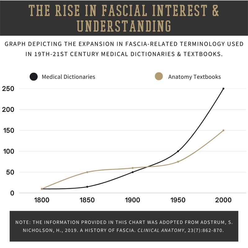
While the revolutionary findings of fascial research are exciting and help to better shape our approach to injury restoration and sport performance, we should remain mindful that there is still an abundance of unknowns regarding the human body and performance optimization. Not only are fascial concepts still largely overlooked in strength and conditioning, but there is still disconnect among experts and governing bodies. For instance, how fascia should be properly defined, what its functions are, and how significant fascia may be in performance are all still being debated and determined.
In this article, I’d like to cover a base understanding of myofascial meridians and how this realization has profoundly influenced my approach to training and my perspective of human movement.
[adsanity align=’aligncenter’ id=11160]
What are Myofascial Meridians?
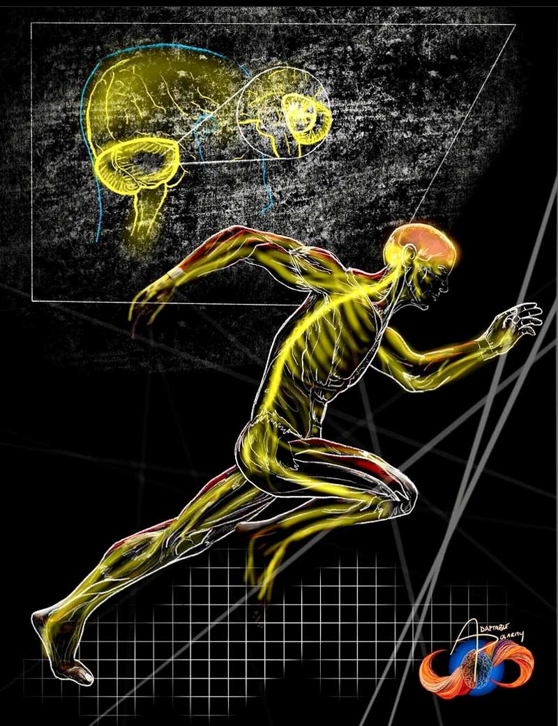
Myofascial meridians are anatomical descriptors that have been broadly defined as continuous bands of fascial tissue spanning across and throughout the body.1 The term meridians, specifically, is one of several terminologies used by prominent modern day fascia researchers. This group of researchers includes:
- Luigi, Antonio, and Carla Stecco
- Robert Schleip
- Jan Wilke
There are other schools of thought, however, that use different terminologies. For instance, another prominent fascia researcher, Tom Myers, uses the term “trains” to classify these fascial vectors. For all intents and purposes, these terms (trains, meridians, lines, and chains) can be viewed as interchangeable and taken to broadly mean the same thing.
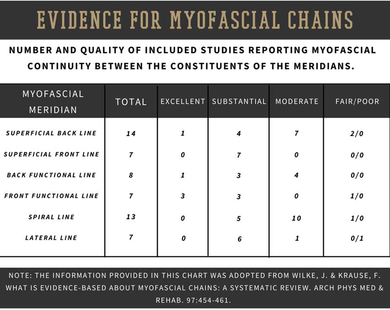
Along with the differences in terminology, there is also disconnect among precisely how many meridians or trains there are in the human body. Tom Myers and the Anatomy Trains organization have stated there are 12 identifiable fascia trains in human anatomy.2 According to an investigative study conducted by Wilke et al.,3 however, they were able to confirm 3 of 6 fascial lines selected form Myers’ original proposed 12. As for the Steccos, along with several other prominent researchers, they typically recognize about 6 meridians in the body.
Although it is a bit unclear as to specifically why there is disconnect among these experts, a part of the difference in numbers may be due to dissection techniques (or skill) and how the tissue is extracted. Additionally, there is still differentiation between tendinous tissue, about which some researchers have different views on what is a part of the tendon proper and what is identified as fascial tissue.
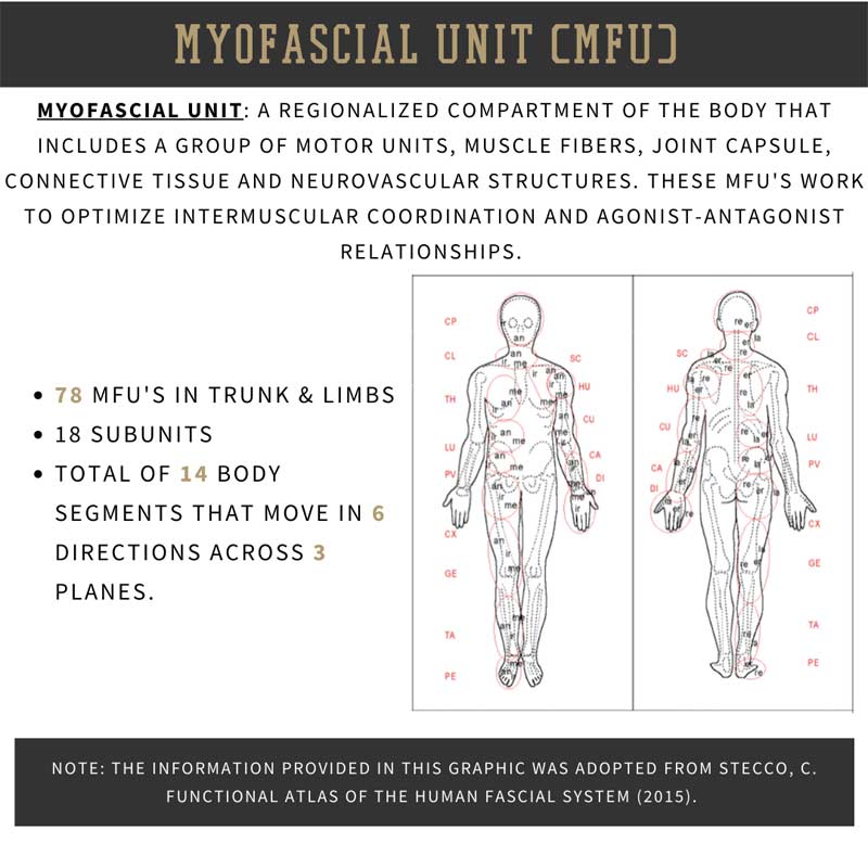
Beyond the meridians themselves, myofascial unit (MFU) is another fascia-specific term that coaches should be adept with. Myofascial units are defined as regionalized compartments of the body that include a group of motor units that activate adjacent muscle fibers that move a body segment in a unidirectional manner.4 This also includes the joint, soft tissue, neurovascular components, and the connecting fascia.4 To simplify that, MFUs represent a localized compartment of the body (i.e., shoulder girdle, posterior hip).
MFUs represent a localized compartment of the body (i.e., shoulder girdle, posterior hip), says @danmode_vhp. Share on XMyofascial units largely speak to the agonist–antagonist relationships, and intermuscular coordination of the muscles and structures in working proximity to coordinate multidirectional movements. And, according to the Steccos, there are 78 identifiable MFUs found in the body. These MFUs can then be broken down and organized into 14 body segments that move in 6 directions across 3 cardinal planes.
I see the myofascial meridians as vectors of kinetic continuity that represent empirical force channels promoting the most efficient pathways for force to be transmitted and directed. These meridians represent lines of chronic stress or critical load vectors whereby the meridians overlay specific regions of anatomy that are activated during common everyday life and sport activities (i.e., walking, twisting, and bending). I can’t reinforce enough that there is no separation between fascia and the musculoskeletal system—everything is working in tandem to produce outcomes.
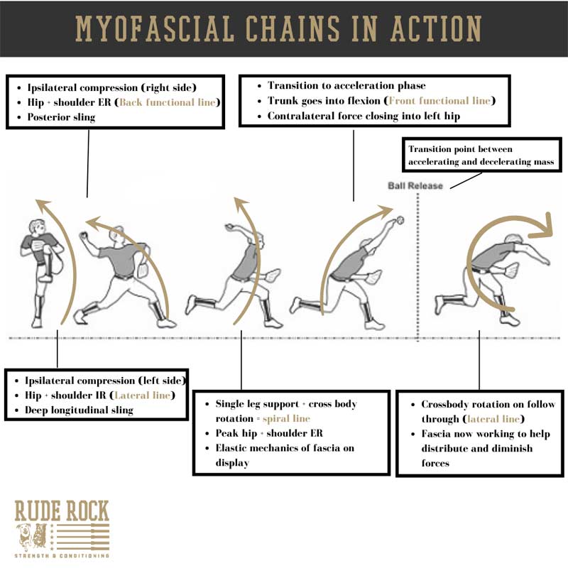
However, when we modify the perspective from which we analyze movement, it can create an invigorating observation. A great example of this is watching a baseball pitcher throw, which is one of the most beautiful displays of human biomechanics. From the fascial point of view, we can see a sophisticated sequence of shifting center of mass, redirecting vectors, and a “dance” between compression and tension throughout the body.
Meridians & Trains > Cardinal Planes
The deeper I investigate into the fascial system, the more I see it as filling in the gaps where conventional anatomy seems to fall short. Moreover, I’m able to recognize the extreme sophistication of human anatomy and movement—and while oversimplification can be effective for introducing concepts, we cannot shy away from the depth of details. A common example of this reductionist nature can be found in the observation of movement through the lens of three cardinal planes. The body, segmentally and collectively, simply does not move in a purely linear fashion—there is rotation and angulation that makes it all seamless.
Applying this to the baseball pitcher above, make note of the anatomical relationships occurring, not only the independent components. Along with the angles and positions of the body, consider how momentum is transferred and the tremendous amounts of torque that are placed on certain joints. While isolated items like glenohumeral internal/external rotation ratios do have significance for a baseball pitcher, I would argue the kinetic function of the anterior/posterior meridians could be more relative to play and performance.
The fascial term for this collective integration is what’s known as biotensegrity. In a nutshell, biotensegrity can be understood as the complex balance of compression and tension forces throughout the body.5 This balancing act is predominantly undertaken by the global fascial net encasing the body, whereby changes in position, speed, or expressions of force alter the localized fascial compartments. Collectively, this distribution of force, although produced by the bones and muscles, is mediated by the global fascial net.
In a nutshell, biotensegrity can be understood as the complex balance of compression and tension forces throughout the body, says @danmode_vhp. Share on X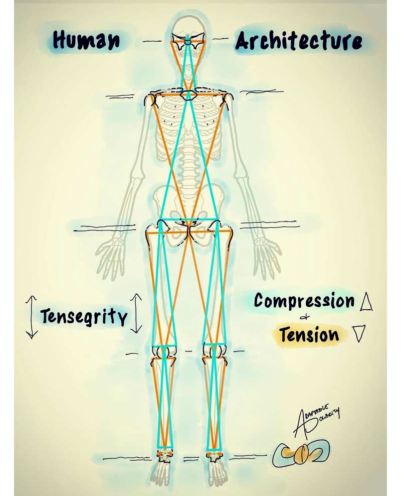
Perceiving the body from the biotensegrity viewpoint as opposed to the three cardinal planes has helped my coaching immensely. The way I observe and analyze movement has become more focused on a global framework, rather than emphasizing the body as being a summation of independent parts. This has helped prevent me from being unnecessarily siloed into one segment or specific joint, instead focusing on understanding the kinetic relationships of the area with the rest of the body. The biotensegrity perspective has also dramatically shifted my approach for exercise selection and training parameters. In a broad sense, this has evolved into more emphasis on global patterns, utilizing more omnidirectional movements, and emphasizing open chain variations with fewer constraints.
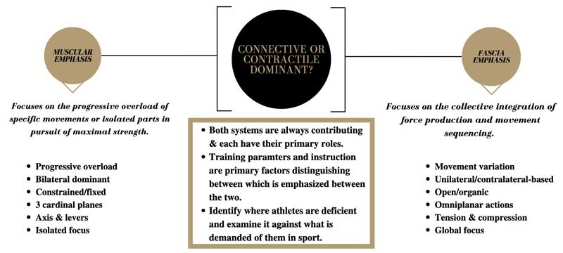
There can be an appropriate time and place for just about anything in training, so again, don’t mistake this for completely doing away with foundational lifts or using isolation exercises. However, these have become much less frequent options for me, and in my opinion do not have as much benefit as we’ve been led to believe. We need to be clear in understanding that the goals of performance training are ultimately to improve the athlete’s ability in sport and minimize the likelihood of injury occurrence.
It should not be an inherent priority to improve numbers on bench, squat, or clean just for the sake of doing so. Rather, we want to look at how we can improve force expression and tolerance across a wide spectrum of positions, vectors, and under varying speeds as they relate to sport.
[adsanity align=’aligncenter’ id=11142]
Aligning Conventional & Contemporary Perspectives
Suggesting that our conventional framework for anatomy and biomechanics is incomplete does not imply we need to tear the whole framework down and start from scratch. But if we have been working from a model that has not been telling us the full story of human biology, I would argue it should put some sense of urgency on us to continuously evaluate our practice. The root of this belief stems from recognizing the process of cadaver research in America, which involves embalming the bodies and using an array of chemicals to preserve the body for research trials and investigation.
The embalming and chemicalization process for cadaver research dramatically changes the biological landscape as it is in living humans. Among these changes, and most prominent for the sake of this article, is the erosion of fascial tissue due to the presence of embalming fluid. As a result, the majority of cadaver research involves bodies that do not have intact fascial tissue, other tissues that have been dehydrated, and drained of most blood volume creating a biological environment that does not properly illustrate the reality. So again, the framework for our understanding of anatomy may be prudent, but it is still not giving us the full scope.
The embalming and chemicalization process for cadaver research dramatically changes the biological landscape as it is in living humans, says @danmode_vhp. Share on XThere will always be disconnect between coaches/practitioners regarding optimal human performance practices and applications: everything from academic background, formative development, and even just geographic location. Fascia concepts are a prominent example of this divide, and understandably so. I will be the first to admit these theories and concepts can be radical to digest, especially for those who have extensive experience in the field.
Nevertheless, I do believe we will be able to establish common ground in the near future. And as more definitive research continues to grow, the more likely we can come to terms with the symbiotic function of both fascial and conventional anatomy. But in the short term, I encourage you to simply suspend your disbelief. Give an honestly open window for being impressionable and see how these concepts make sense to you and apply to your setting.
Since you’re here…
…we have a small favor to ask. More people are reading SimpliFaster than ever, and each week we bring you compelling content from coaches, sport scientists, and physiotherapists who are devoted to building better athletes. Please take a moment to share the articles on social media, engage the authors with questions and comments below, and link to articles when appropriate if you have a blog or participate on forums of related topics. — SF
References
1. Findley, T.; Chaudry, H.; Stecco, A.; and Roman, M. “Fascia research: A narrative review.” J Bodywork & Mvmt Thera. 2012;16, 67-75.
2. Myers, T. Anatomy Trains: Myofascial meridians for manual and movement therapists. 2ND ed. Churchill Livingstone, Edinburgh, 2009.
3. Wilke, J.; Krause, F.; Vogt, L.; and Banzer, W. “What is evidence-based about myofascial chains: a systematic review.” Arch Phys Med & Rehab. 2016;97:454-461.
4. Maas, H.; Sandercock, TG. “Force transmission between synergistic skeletal muscles through connective tissue linkages.” J Biomed and Biotech. 2010.
5. Scarr, G. Biotensegrity: The structural basis of life. United Kingdom, Handspring Publishers, 2014.
6. Adstrum, S. Nicholson, H., 2019. A history of fascia. Clinical Anatomy, 23(7):862-870.
7. Krause, F. Wilke, J. Vogt, L. Banzer, W., 2016. Intermuscular force transmission along myofascial chains: a systematic review. J Anat., 228:910-918.
8. Wilke, J. Krause, F. Vogt, L. Banzer, W., 2016. What is evidence-based about myofascial chains: a systematic review. Arch Phys Med & Rehab, 97:454-461.
9. Myers, T. Anatomy Trains: Myofascial meridians for manual and movement therapists- 2ND ed. Churchill Livingstone, Edinburgh, 2009.


