A Protocol for Two-Dimensional Running Gait Analysis

Summary
While 3D motion capture is the gold standard for movement analysis, if your clinic, club, or team doesn’t have the necessary equipment or personnel, you may conduct running biomechanics evaluations using a 2D motion analysis system. This article presents a plan using 2D motion analysis to help identify biomechanical factors related to common injuries in…
With a reported 2 million people running in the U.K.1 and an estimated 10 million in the U.S.,2 you can inarguably say that running fits as snugly into modern life as an eager foot into a pair of new sneakers. Apart from being a popular recreational activity, running also forms the very core of so many sports at the competitive level.
With such an exponential increase in running activity all over the world, a number of studies have reported an increase in running-related injuries at both competitive and recreational levels. The annual rate of running-related injuries ranges from 24-65%, with the most commonly injured joint being the knee. Among runners training for a marathon, the injury rate has been reported as high as 90%.3,4
These injuries can generally occur secondary to four main factors:
- Musculoskeletal impairment.5
- Extrinsic causes such as running surface or footwear.6
- Training error.7
- Faulty running mechanics.8
However, with advancements in measurement technology, our ability to capture running mechanics as part of a routine clinical exam has increased, as well as our ability to integrate the data provided by a running gait analysis into the clinical decision-making process.
As the head of development for a motion analysis software called GaitON, I will share a standardized approach we use to assess running mechanics using 2D motion analysis.
The Equipment for a Running Gait Analysis
It is a universally accepted fact that a three-dimensional (3D) motion capture is the gold standard for movement analysis, providing the most accurate and comprehensive means to assess running mechanics. However, due to its high cost and lack of portability, most clinics don’t have the necessary equipment or personnel to use such systems, thereby limiting its use in general practice.
A 2D motion analysis system is affordable, and offers a high degree of portability. Share on XA 2D motion analysis system, on the other hand, is not only more affordable, but also offers a higher degree of portability to a clinician. Having said that, it is absolutely essential that the analysis protocols used in a 2D motion analysis system are reliable and validated against the gold standard (3D motion analysis).
Hence, the objective of this article is to provide a framework for a systematic running biomechanics analysis that you can conduct using a 2D motion analysis system. Although some of the proposed variables of interest will have an impact on running performance, the primary focus of this analysis plan is to identify biomechanical factors related to common injuries in a runner.
The Running Surface: Treadmill vs. Overground Running
Although a few studies have identified small differences in treadmill running when compared with overground running, these differences have mostly been associated with muscle activation patterns and joint forces.9 In general, kinematic patterns during treadmill running are very similar to those observed during overground running.10
As such, performing a video-based analysis of joint kinematics during treadmill running can provide valuable insight into running kinematics during overground running and is more practical, considering the space availability in clinics.
What is more important is that the runner is comfortable with the environment and the analysis is done at a comfortable running speed.
Views of Motion Capture
Typically, any good motion analyzer supports up to two digital cameras and provides data from the anterior/posterior views and the right/left lateral views of a runner. Each view provides information about different parameters that aid in the assessment of the running biomechanics.
How Detailed Should the Analysis Be?
Understanding the running gait cycle is imperative for proper clinical gait analysis and communication with runners. Although modern-daymotion analyzers provide a complete breakdown of the entire running gait cycle, analyzing the running form at a few key moments is generally sufficient. These key moments include the positions of:
- Initial Contact: Instant at which the foot first makes contact with the ground.
- Mid Stance: Instant when the body’s center of mass (COM) is directly over the foot.
- Toe Off: Instant at which the toe leaves the ground.
Analyzing the Lateral View
The sagittal plane body postures have been known to provide insights into the ground reaction forces and the joint kinetics acting on the runner. While analyzing this view, it is helpful to develop a systematic top-down or bottom-up approach, with particular attention paid to the initial contact and mid stance positions.
Initial Contact
- Foot Inclination Angle
The foot inclination angle is the angle between the running surface and the sole of the runner’s shoe, and it determines the foot-strike pattern of a runner. An increased value of this angle leads to an increase in peak vertical ground reaction force, braking impulse, knee extensor moment, and negative work performed by the knee extensors, all of which have been associated with various running-related injuries.10
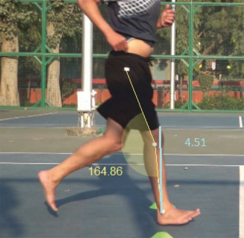
- Leg Inclination Angle
The leg inclination angle is the angle of the midline of the leg and the true vertical. (Image 1) The magnitude of this angle contributes to the amount of loading that occurs to the tibia during running. Reduction of stride length is a very common solution suggested by clinicians to reduce excessive leg inclination angles, thereby lowering the risk of tibial stress injuries.
- Knee Flexion Angle
The knee flexion angle is measured as the angle between the midline of the thigh and the midline of the leg. (Image 1) Knee flexion angles near 20 degrees are generally recommended.
Mid Stance
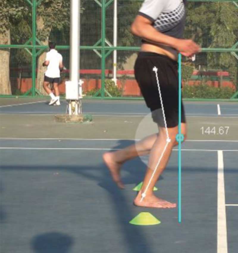
- Knee Flexion Angle
This angle is highly predictive of peak patellofemoral joint force, such that peak force increases as knee flexion angle increases.11(Image 2) Although the value of this angle (like all joint kinematic values) will vary with speed, knee flexion angles near 40-45 degrees are generally recommended.
- Ankle Dorsiflexion Angle
Assessing peak ankle dorsiflexion during stance may provide insights into calf muscle load and Achilles tendon strain.
Toe Off
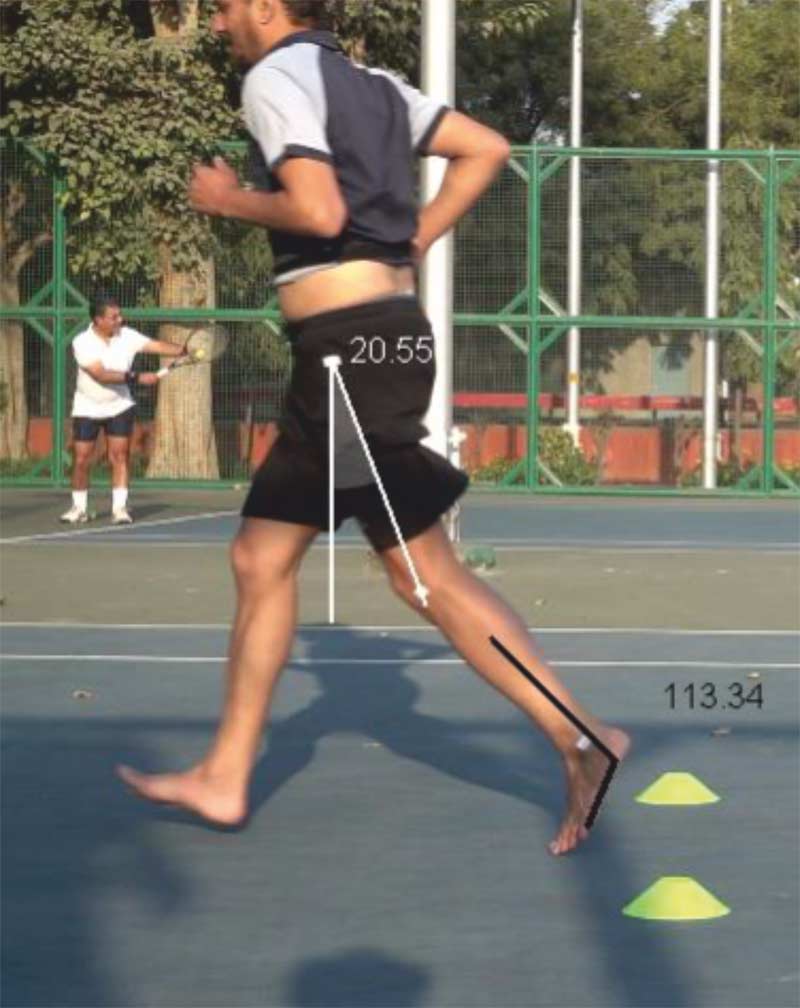
- Hip Extension
Although this is still a topic of ongoing research, hip extension is certainly an important measurement to assess, especially in those with low back or hip pain and in the postpartum running population. (Image 3)
- Ankle Plantarflexion
Ankle plantarflexion is an essential parameter for assessing restricted ankle motion, restricted great toe motion, or weakness of the calf muscles, and the resulting compensations that may lead to Achilles tendonitis or plantar fasciitis, and may even affect the knee, hip, or low back. (Image 3)
Analyzing the Posterior View
The posterior view best captures the runner in the mid-stance position of the gait cycle.
Mid Stance
- Trunk Side Bending
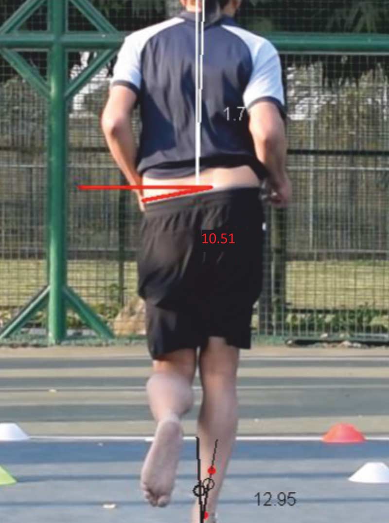
Trunk side bend, defined as midline of the trunk relative to the vertical, can demonstrate motion in either the ipsilateral or contralateral directions. Increased ipsilateral trunk lean is found in runners with both patellofemoral pain and iliotibial band syndrome,12 often associated with increased motion of the pelvis in the frontal plane. (Image 4)
- Lateral Pelvic Drop
Lateral pelvic drop is defined by a line connecting the iliac crests relative to the horizontal. Keep gender difference in mind while assessing this parameter, as the amount of pelvic drop is usually higher in females. (Image 4)
- Rearfoot Eversion
Rearfoot eversion is defined by the midline of the heel relative to the midline of the lower leg. (Figure 4). Several studies have linked excessive heel eversion to various running injuries, such as tibial stress fractures, patellofemoral pain, and Achilles tendinopathy.13 Furthermore, it has been suggested that runners with excessive calcaneal eversion be prescribed orthotics,14or a higher level of support shoes.
How to Ensure the Accuracy of Measurements
The accuracy of the results given by any motion system is dependent on three factors: marker placement, system reliability, and reliability of reference values.
Marker Placement
Although you can do the measurement of joint angles without any markers, research suggests that placing markers over anatomical landmarks improves the reliability of the analysis. However, if you place the markers incorrectly, it can introduce further errors into measurement.
The (proper) placement of markers over anatomical landmarks improves the reliability of analysis. Share on XSystem Reliability
While doing our competitor research for motion analysis software, we came across a lot of freeware in the market (along with a few paid ones too) that don’t have built-in protocols for motion analysis, thereby forcing the user to measure these angles manually. Such practices not only reduce the reliability of the results (drastically!), but also lead to increased analysis time for clinicians.
Hence, if reliability is critical to the analysis, you should use a software that provides this facility.
Reliability of Reference Values
Establishing reference values for comparison helps clinicians evaluate what is normal mechanics and what is not, thereby allowing them to make targeted interventions. Again, a lot of software, like GaitON, have built-in reference values that assist clinicians by providing a complete motion analysis report.
A Starting Template
I touched upon a few points that can help clinicians perform a running gait evaluation. Although these parameters are not the only ones that you can measure during a running gait analysis, these may certainly serve as a template for a systematic evaluation plan.
If there is any suggestions or discussions you would like to make, you can write to me at [email protected]
References
- Finn, A., 2013. “Why we love to run.”[Online]
Available at: https://www.theguardian.com/lifeandstyle/the-running-blog/2013/feb/05/why-we-love-to-run - USA Track & Field, n.d. “Long Distance Running – State of the Sport.”[Online]
Available at: http://www.usatf.org/news/specialReports/2003LDRStateOfTheSport.asp - van Gent, R.N. et al., 2007. “Incidence and determinants of lower extremity running injuries in long distance runners: a systematic review.” British Journal of Sports Medicine. 41(8).
- Macera, C. et al., 1989. “Predicting lower-extremity injuries among habitual runners.” Archives of Internal Medicine.149(11): 2565-8.
- Dierks, T., Manal, K., Hamill, J. & Davis, I., 2008. “Proximal and distal influences on hip and knee kinematics in runners with patellofemoral pain during a prolonged run.” The Journal ofOrthopaedic & Sports Physical Therapy. 38(8): 448-56.
- Taunton, J. et al., 2003. “A prospective study of running injuries: the Vancouver Sun Run ‘In Training’ clinics.” British Journal of Sports Medicine. 37(3).
- Lysholm, J. & Wiklander, J., 1987. “Injuries in runners.” American Journal of Sports Medicine. 15(2): 168-71.
- Nakagawa, T., Moriya, É., Maciel, C. & Serrão, A., 2012. “Frontal plane biomechanics in males and females with and without patellofemoral pain.” Medicine and Science in Sports and Exercise. 44(9): 1747-55.
- Sinclair, J. et al., 2013. “Three-dimensional kinematic comparison of treadmill and overground running.” Sports Biomechanics. 12(3): 272-82.
- Fellin, R., Manal, K. & Davis, I., 2010. “Comparison of lower extremity kinematic curves during overground and treadmill running.” Journal of Applied Biomechanics. 26(4): 407-14.
- Wille, C. et al., 2014. “Ability of sagittal kinematic variables to estimate ground reaction forces and joint kinetics in running.” Journal ofOrthopaedic & Sports Physical Therapy. 44(10): 825-30.
- Noehren, B. et al., 2012. “Proximal and distal kinematics in female runners with patellofemoral pain.” Clinical Biomechanics. 27(4): 366-71.
- Barton, C., Bonanno, D., Levinger, P. & Menz, H., 2010. “Foot and ankle characteristics in patellofemoral pain syndrome: a case control and reliability study.” Journal ofOrthopaedic & Sports Physical Therapy. 40(5): 286-96.
- Kannus, V., 1992. “Evaluation of abnormal biomechanics of the foot and ankle in athletes.” British Journal of Sports Medicine. 26(2): 83-89.
